Pathophysiology of Myelodysplastic Syndromes (MDS)
The underlying pathophysiology of MDS is complex and heterogeneous, and may develop over decades, resulting in progressive dysplasiaGerming U et al. Ann Hematol. 2023;102:311-321. Sekeres MA, Taylor J. JAMA. 2022;328:872-880.
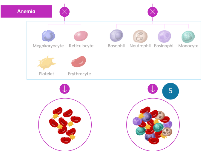
MDS is a spectrum of neoplastic bone marrow failure disorders characterized by ineffective hematopoiesis, resulting in morphologic dysplasia and various degrees of cytopenia.
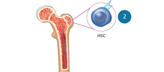
- MDS is a clonal disorder that arises from the expansion of hematopoietic stem cells (HSCs)Sekeres MA, Taylor J. JAMA. 2022;328:872-880. Hoff FW, Madanat YF. Cells. 2023;12:627.
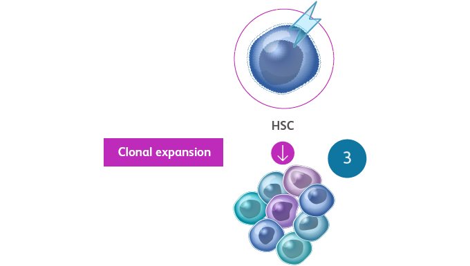
- The somatic mutations that drive MDS confer selective survival advantage over wild-type cells, and the enhanced self-renewal leads to the accumulation of clonal hematopoiesis over time, resulting in abnormal progenitor and precursor cellsSekeres MA, Taylor J. JAMA. 2022;328:872-880. Hoff FW, Madanat YF. Cells. 2023;12:627.
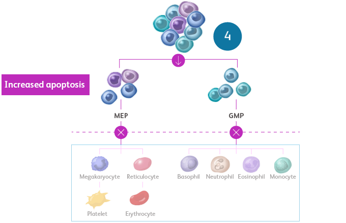
- Terminal maturation is impaired, leading to increased apoptosis of differentiating cells and, thus, peripheral cytopeniasSekeres MA, Taylor J. JAMA. 2022;328:872-880. Hoff FW, Madanat YF. Cells. 2023;12:627. Sperling AS et al. Nat Rev Cancer. 2017;17:5-19.
- In 30% of cases, MDS can progress to acute myeloid leukemia (AML) because of the accumulation of additional mutationsHoff FW, Madanat YF. Cells. 2023;12:627.
Ineffective hematopoiesis in MDSSekeres MA, Taylor J. JAMA. 2022;328:872-880. Hellström-Lindberg E et al. Haematologica. 2020;105:1765-1779. Adès L et al. Lancet. 2014;383:2239-2252.

HSC, hematopoietic stem cell.
An initiating driver mutation occurs in an HSC, giving rise to clonal hematopoiesis of mutant stem cells and abnormal progenitor and precursor cells.Sekeres MA, Taylor J. JAMA. 2022;328:872-880.
![]()
Clonal hematopoiesis of indeterminate potential (CHIP) is defined by the presence of a somatic mutation in a myeloid neoplasm driver genes or a non-MDS-defining clonal cytogenetic aberration, in a patient with no known myeloid neoplasm and no unexplained cytopenia.Hasserjian RP et al. Virchows Arch. 2023;482:39-51.
Multiple chromosomal abnormalities and genetic mutations have been identified to cause MDSVeiga CB et al. Cancers (Basel). 2021;13:1968.
To learn more, view the tabs below. Each tab contains helpful information.
Most common cytogenetic mutations in MDSHoff FW, Madanat YF. Cells. 2023;12:627. Saygin C, Godley LA. Cancers (Basel). 2021;13:3380.

del, deletion.
- Approximately half of patients with MDS harbor chromosomal abnormalities affecting copy number alteration (eg, deletion, monosomy, or trisomy) or, more rarely, leading to a structural change (eg, balanced translocation or inversion)Hoff FW, Madanat YF. Cells. 2023;12:627.
- The most common chromosomal abnormality is del(5q), which is present in up to 15% of patientsHoff FW, Madanat YF. Cells. 2023;12:627.
- Up to 30% of patients with MDS exhibit a complex karyotype (≥ 3 cytogenetic abnormalities), which is associated with a higher risk of progression to AML and a very poor prognosisHoff FW, Madanat YF. Cells. 2023;12:627.
Landscape of somatic mutations in MDSHoff FW, Madanat YF. Cells. 2023;12:627. Tobiasson M, Kittang AO. J Intern Med. 2019;286:41-62.
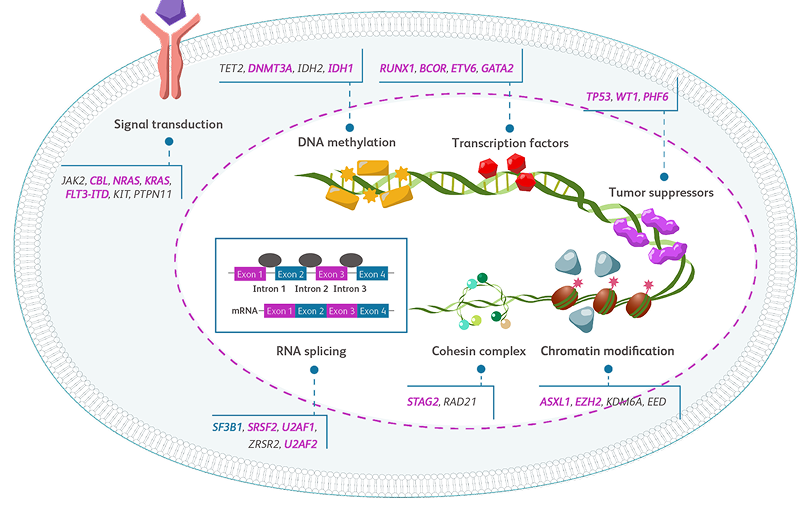
Adapted with permission from J Intern Med.Tobiasson M, Kittang AO. J Intern Med. 2019;286:41-62.
DNA, deoxyribonucleic acid; mRNA, messenger RNA; RNA, ribonucleic acid.
- Whole-exome sequencing technologies have enabled detection of recurrent somatic mutations in > 50 genes in up to 90% of MDS casesSaygin C, Godley LA. Cancers (Basel). 2021;13:3380.
- Target driver gene mutations are involved in a variety of functional pathways, as shown belowSaygin C, Godley LA. Cancers (Basel). 2021;13:3380.
Frequency of driver gene mutations in MDSHoff FW, Madanat YF. Cells. 2023;12:627. Nazha A, eds. Diagnosis and Management of Myelodysplastic Syndromes: A Clinical Guide. Springer; 2020.

Adapted with permission from Springer.Nazha A, eds. Diagnosis and Management of Myelodysplastic Syndromes: A Clinical Guide. Springer; 2020.
Recurrently mutated genes in MDS, categorized according to biological function and mutation frequency. Circle size correlates with mutation frequency; light-colored halos indicate the upper limit of frequency. Mutations that confer an IPSS-R—independent negative effect are colored in blue; mutations with no clear independent effect are displayed as gray/light gray circles. Only SF3B1 mutations are associated with a favorable prognosis (purple).Nazha A, eds. Diagnosis and Management of Myelodysplastic Syndromes: A Clinical Guide. Springer; 2020.
DNA, deoxyribonucleic acid; IPSS-R, Revised International Prognostic Scoring System; RNA, ribonucleic acid.
Learn more

